Microscopy
Micro-what?
You may have heard the word microscope, which is that thing that scientists use when they want to look science-y. (And when they want to look at really small stuff. Which, depending on the scientist, might be all day, every day). It’s basically a really fancy, powerful magnifying glass. Microscopy is just the use of a microscope.
To talk about cells, we really have to mention microscopes. That’s because cells are microscopic, so we need a microscope to see them. Most things we know about cells we know, at least in part, because of microscopy.
To talk about cells, we really have to mention microscopes. That’s because cells are microscopic, so we need a microscope to see them. Most things we know about cells we know, at least in part, because of microscopy.
Types of Microscopes
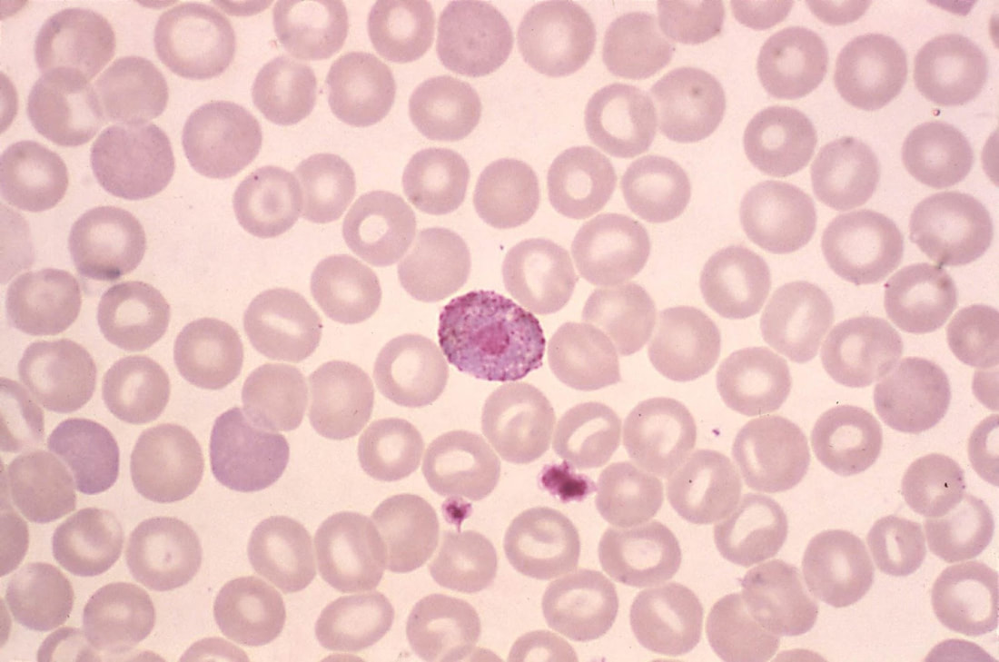
Regular light microscopes—the ones you’re probably most familiar with—can allow us to see up to about 400 times the size of an object. They might take an image like this:
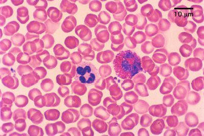
Blood under a light microscope
There are also other kinds of microscopes, like fluorescence microscopes, which might take an image like this:
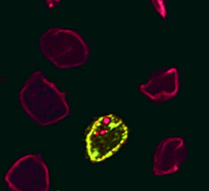
Blood under a fluorescence microscope
Another type of microscope is the electron microscope, which can magnify up to 500,000 times. They might take an image like this:
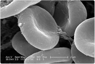
Blood under an electron microscope
There are also many other types of microscopes out there. The only one you need to know the details of for this class is the standard light microscope.
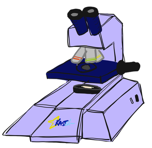
Standard Light Microscope
This is a standard light microscope, like one that you might find in just about any biology classroom or lab (including the labs that real biologists use) around the world:
The key parts of this are the eyepiece (the part you look through with your eyes, so that you can actually see what you’re looking at--it also magnifies somewhat), the light source (which makes things bright, so that you can see), and this part...:
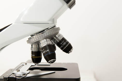
...which is called the objective. This is where most of the magnification happens.
Most microscopes, including this one, have multiple objectives in different magnifications (usually 4x to 40x, for a total magnification, when you factor in the eyepiece, of 40x to 400x). Larger objectives give more magnification, so that you can see the smallest stuff. Having multiple objectives gives the viewer control over how much he or she wants to zoom in. You can look at stuff in “wide view” (for example, using a 4x objective) to get a more complete picture of what something looks like, and “super-close up” (for example, using a 40x objective) to get in super close on the details.
The logic behind which one to use is basically the same as how you would decide whether to take a picture of someone where you can see their whole body versus one that’s zoomed in really close on their nose. Both could be interesting, but they have very different purposes. If, for example, you’re trying to see if a person has freckles, the really zoomed in one might be more helpful.
Light microscopes are pretty easy to use, once you know how to do it. As such, watching someone use a light microscope can be pretty boring. If you have the ability to use a microscope in person, it can be a lot of fun to look at all sorts of different everyday objects just to see what you can see, and to learn about the microscopic structure of these everyday objects. STEM (Science, Technology, Engineering, and Mathematics) fairs and camps are a great place to do this. Schools and community centers also often have microscopes, and some relatively cheap ones designed for kids to explore with can be purchased in stores or online.
This video gives a great overview of using a microscope:
Neat, right?
Looking At Stuff Under A Microscope
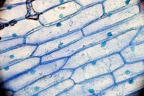
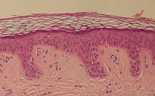
So, what does stuff look like under a microscope? It’s a little bit tricky to show you on a computer screen exactly what something looks like at a certain magnification under a microscope, because pictures change size depending on the size of your screen and how much you are zoomed in. But here are some cool pictures of stuff under microscopes, just because it’s interesting. Note that the colors come from how we stain stuff to see it better; really, this close up, most stuff is just various shades of clear:
|
|
|
|
Blood
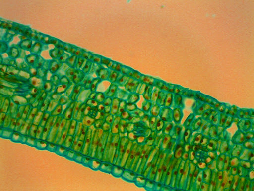
Leaf
|
Onion
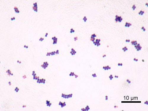
Staphylococcus aureus, a common type of bacteria (Staph infection)
|
Skin
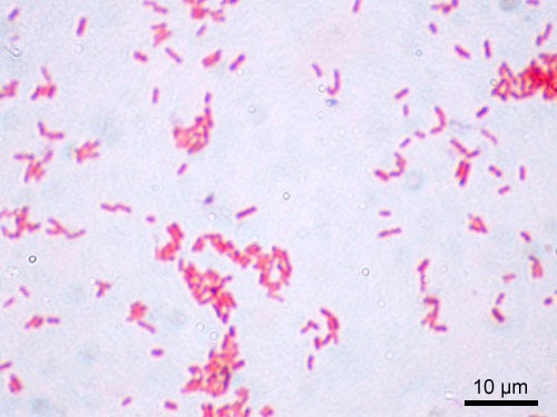
Escheria coli, another common type of bacteria (E. coli)
|
Feel free to search the web (using all standard precautions for searching the web, of course) for pictures of whatever interesting thing strikes your fancy under a microscope.
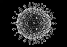
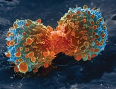
Just for fun, here are some pictures of stuff under a scanning electron microscope, which is a super powerful kind of microscope that lets you see a lot of the really cool close-up 3D details of stuff. The colors are added in post-processing for artistic effect:
|
|
|
Flu virus
|
Cancer Cell Dividing
|
Summary
You should understand:
- How to use a light microscope.
Learning Activity
Content contributors: Emma Moulton and Emily Zhang